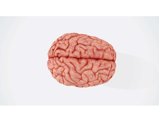
What does anger look like in the brain? And how can that knowledge inform therapy and personal growth? This article dives into these questions, and more.
Jump to section:
Introduction
For aeons, human beings have recognised the physical sensations and mental and emotional effects associated with anger. With the advent of fMRI and other brain-imaging technology, however, we have also been able to establish what parts of the brain “light up” or activate during an escalation of emotions into anger. In this article, we outline the physiological path through the body-mind that anger takes when we first detect a threat of some sort. As we proceed through this discussion, there are multiple parts involved, including names for neurotransmitters and brain parts that are somewhat technical. The main thing to focus on, though, is the interplay between the part of us that just reacts emotionally – protectively – to the threat and the part of ourselves that can take the time to reason out what, if any, protective action is necessary. Tip: To explore common myths about anger and anger management, read Busting 10 Common Myths About Anger.
Amygdala: The seat of emotions
Brain experts are fond of talking about two almond-shaped structures in our brains, called the amygdala, as the place where emotions begin. It is located in the back of the brain: that is, the “old” brain, present before modern human beings began serious development of their executive pre-frontal cortex in the front of the brain (see Figure 1). The amygdala’s job is to identify threats to our wellbeing, and sound the alarm when any such perceived threats are identified, motivating us to take protective action. The problem for dealing well with anger, as we will see, is that the amygdala is hard-wired to respond immediately: before the “voice” of reason, logic, and judgment (the pre-frontal cortex) has time to adequately consider what’s happening.
Preparing for the fight
As a person becomes angry, the body muscles tense up. Neurotransmitters known as catecholamines are released, giving the person a burst of energy that can last up to several minutes: hence, our desire to take that protective action right now. At the same time, physical changes in the body begin to prepare the person for the fight; accelerating heart rate, increasing blood pressure and rate of breathing, and flushed face are all part of the increased blood flow entering a person’s limbs and extremities in preparation for physical action.
A person’s attention narrows, eventually becoming fixed on the target of the anger, with difficulty noticing anything else. More neurotransmitters and hormones arrive on the scene, among these adrenalin and noradrenalin. Of course, bodily processes such as digestion work differently so that vital energy can be preserved for the fight (MentalHelp.net, 2020) (see Figure 2).
Figure 1: The amygdala and pre-frontal cortex

(Cashcoach.io, 2020)
Figure 2: What happens to your body when you are angry

(Adapted from Stosny, n.d.)
We know that if an angry or stress response continues to occur in the body, we eventually end up with adrenal exhaustion. Figure 3 traces this route from the amygdala to the adrenal glands.
Figure 3: How anger affects your brain and body

(Adapted from Gray, 2018)
The adult in the room: The pre-frontal cortex
While there are certainly myriad situations these days in which we do need to act protectively and immediately (think dodging a car careening around the corner as you finish crossing the street or grabbing a small child just before they tumble into a rapidly flowing stream), the reality of modern life is that the threats tend to be more psychological than physical. Perhaps our partner criticises us unjustly, we get a huge bill unfairly and unexpectedly, or we attend an awards ceremony where the top prize goes to a competitor whose work we did not think was very good, or at least not as high-quality as our own. In these cases, little is accomplished through unsheathing our metaphorical sword (that is: the amygdala letting rip).
Enter the part of ourselves that can put the brakes on that all-protective but somewhat Neanderthal amygdala: the pre-frontal cortex (PFC). The PFC is the part of the brain that enacts reasoned judgment, logic, and well-thought-out responses. The PFC, for example, would be unlikely to contradict the urging of the amygdala that got you out of the way of the oncoming car. But, when it’s properly engaged, it might very well persuade you to hold fire on impulsive angry retorts to a critical boss. Many schools of therapy are now following the lead of dialectical behaviour therapy (DBT) in recognising that amygdala-originating emotions not reined in by the PFC create a dangerous situation of emotional dysregulation for some clients (for example: those who are suicidal or with eating disorders). To regulate ourselves emotionally is to recruit the PFC in its built-for-purpose executive role to keep things on a more even keel (MentalHelp.net, 2020). We can’t, of course, control what happens to us, but we can work at controlling our reactions to events.
Research on the activation of different brain areas
Here we note research by Klimecki, Sander, & Vuilleumier (2018) using fMRI neuroimaging to study whether people use different parts of their brain to register anger (an amygdala function) than to regulate punishment-related behaviours (more of a PFC function). The researchers observed participants playing an economic interaction paradigm called “Inequality Game” (IG). The game has been validated for its capacity to elicit anger through the competitive behaviour of an unfair (versus fair) other and also to promote punishment behaviour. Results showed that different parts of the participants’ brains lit up, or activated, when they felt angry about an unfair other’s playing (when the participant was in the second, low-power phase of the experiment) versus when those same participants were engaged in decisions about whether or not to punish the unfair other in the third part of the experiment when they were in the high-power position (the first phase for participants had them in a high-power position with their competitors in order to establish baseline tendencies toward cooperation or competitiveness).
In addition to the fMRI, the researchers assessed the effects of the unfair other’s behaviour on participants’ feelings through questionnaire-based measures of participants’ emotions. The extent to which participants engaged in cooperative as opposed to competitive economic choices towards the unfair other in the high-power phase after anger provocation (compared to their baseline score) served to assess the degree of participants’ punishment inhibition.
In their discussion, Klimecki and colleagues assert that this study extended previous research by dissociating the brain activity related to anger experience during provocation from subsequent regulations of punishment behaviour. Anger-related brain activations were present in the amygdala and the related superior temporal sulcus (STS) region with amygdala activations related to emotion processing. Meanwhile, stronger activations in the dorsolateral prefrontal cortex (DLPFC) and related anterior cingulate cortex (ACC) when seeing the unfair other predicted the inhibition of aggressive behaviour (i.e., less punishment) in the subsequent phase.
These two interconnected regions are important for conflict resolution, emotion regulation, and the integration of motivational information for guiding goal-relevant decisions. These latter activations emphasised the importance of emotion control during the provocation phase in guiding subsequent punishment inhibition. The findings provide new insights on functional mechanisms of aggression regulation that have important implications for effective clinical interventions in populations with anger or aggression issues (Klimecki et al, 2018).
Thought for reflection
Think of a time in your life when you were seriously provoked toward anger. Which part of your brain ultimately came to the fore in your expression of that provocation: your amygdala or your PFC? In general, what’s your sense of how well regulated your amygdala is by your PFC? What’s one step you could take today to improve your self-regulation?
Summary
For our purposes, it’s affirming to know that neuroimaging research is confirming what we have long suspected: that anger “happens” in a different part of the brain than the part of the brain that decides how to react to the incident provoking the anger. Therefore, our work with clients can help them to develop the parts (the PFC and related regions) which are designed to reason out whether an angry response – “punishment” in the study just cited – is warranted or not.
Key takeaways
- When we perceive a threat, we begin to get angry, and neurotransmitters are released causing both physical and attentional changes to help prepare us to deal with the threat.
- The amygdala in the older part of our brain is tasked with identifying threats to our wellbeing. It is the part that sounds the alarm when any perceived threats are identified, motivating us to take protective action.
- The PFC is the executive (newer) part of the brain that enacts reasoned judgment, logic, and well-thought-out responses; its job is to rein in emotions originating in the amygdala in order to regulate us emotionally.
- Recent research has used fMRI neuroimaging to empirically demonstrate that people use different parts of their brain to register anger (an amygdala function) than to regulate punishment-related behaviours (more of a PFC function).
References
- Cashcoach.io. (2020). Image of amygdala and pre-frontal cortex. Novamoney.com Retrieved on 12 April, 2021, from: https://novamoney.com/images/uploads/4_brain.jpg
- Gray, J. (2018). How anger affects your brain and body. Anger, nursing tips, health guide. Retrieved on 12 April, 2021, from: https://www.nicabm.com/sample/nicabm-infographic-anger/
- Klimecki, O.M., Sander, D., & Vuilleumier, P. (2018). Distinct brain areas involved in anger versus punishment during social interactions. Scientific Reports (2018) 8: 10556/ DOIL10.1038/s41598-018-28863-3. Retrieved on 12 April, 2021, from: https://www.nature.com/srep/
- Mentalhelp.net. (2020). Physiology of anger. American Addiction Centers. Retrieved on 12 April, 2021, from: https://www.mentalhelp.net/anger/physiology/
- Stosny, S. (n.d.). What happens to your body when you are angry (image). Microsoft Bing Images. Retrieved on 12 April, 2021, from: http://capacitybuildingdevelopment.blogspot.com/2016/06/anger-management-isnt-as-difficult-as.html


